Digestive System of a Dog
| ✅ Paper Type: Free Essay | ✅ Subject: Biology |
| ✅ Wordcount: 3518 words | ✅ Published: 23 Sep 2019 |
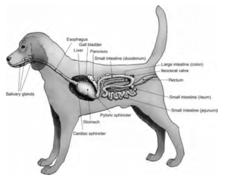 This essay is going to explain the digestive system of a dog, focusing on how the structure aids the function. The digestive system consists of a collection of organs that all have a variety of roles in order to break down food and absorb nutrients. Figure 1 shows the gastrointestinal (GI) tract of a dog which is relatively short. It consists of two groups of organs: the mouth, most of the pharynx, oesophagus, stomach, and intestines, and the accessory digestive organs: teeth, tongue, salivary glands, liver, gallbladder and pancreas (Hume et al, 1995: page 55) (Grabowski et al, 2000: page 818).
Figure 1 (Case et al, 2011)
Even though they bolt their food, little chewing does happen in order for digestion to begin. 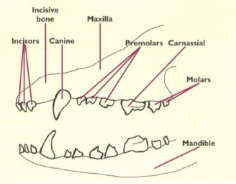 Since Canis lupus familiaris (dogs) are carnivores, their teeth have adapted to enable them to chew and tear meat and they have 42 different structured teeth, shown in figure 2, which allow this function to happen (Jacobs, 2005). The incisors and canines, which are located at the front of the mouth, allow the food to be broken down into smaller molecules before entering the stomach, due to the sharp points (Aspinall, 2014: page 95). The tongue is also able to aid with this since it is composed of bundles of striated muscle, making it strong, and the cranial end is very mobile, so it can shape the food into a bolus making it easier to be chewed and travel down the oesophagus (Aspinall, 2014: page 95). Even though food is not in the dogs mouth a long time, so saliva cannot have much effect, it does help with this process as it lubricates the food aiding with swallowing (Michell et al, 1989: page 90). Paired salivary glands secrete saliva but since this contains no enzymes, rather than breaking down food in the mouth, the main role of saliva in dogs is to help kill bacteria minimising the amount that gets into the stomach (Cowell, 2017).
Figure 2 (Aspinall, 2014)
Stomach
Once the food has passed through the pharynx and oesophagus, it then enters the stomach via the cardiac sphincter. In canines, digestion begins in the stomach where food is broken down into smaller molecules and passed through the GI tract. This is a large muscular bag which stores food temporarily and is the site at which food is converted into a semi – liquid; chyme (Grabowski et al, 2000: page 833). It is also lined with gastric folds and is where acid and enzymes are produced to aid with further digestion.
Figure 3 (Washington State University College of Veterinary Medicine, 2019)
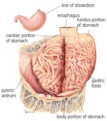 The stomach consists of four regions which are shown on figure 3. The upper cardiac region is at the top of the stomach, surrounding the cardiac sphincter, the fundus is an air-filled section, the corpus is the body of the stomach where the food is stored, and the pyloric region is at the bottom of the stomach connecting to the duodenum. The pyloric region consists of two parts: the pyloric antrum and the pyloric canal (Grabowski et al, 2000: page 833). Pyloric antrum, which is connected to the body of the stomach, contains G cells which secretes the hormone gastrin. The stomach is stimulated by this hormone to release gastric acid. This hormone has two main function: breaking down proteins into amino acids and disinfecting, by killing most of the bacteria which is entered with food (Society for Endocrinology, 2011). After this process, the partially digested food enters the duodenum via the pyloric canal. Dogs eat much quicker than the food can be digested, since liquids can pass through the stomach in half an hour and solids or fatty foods take over four hours (Aspinall, 2014: page 96). As the stomach is a larger storage area at the upper part of the GI tract, it means the intestines can be supplied with partially digested food at an optimised rate whilst the rest is still being stored (Hove et al, 2010: page 578).
The mucosa, a layer of the GI tract wall, lies in large folds when the stomach is empty (Grabowski et al, 2000: page 820). This allows a lot of food to be stored at one time before being slowly released into the intestines, as they expand to accommodate large meals. Due to these folds, which can be seen in figure 3, there is space for more food than can be digested to be stored at one time, giving time for the acid to break down proteins and kill the bacteria before being digested further. This is important in dogs as they eat their food quicker than it leaves the stomach, aiding with the function to serve as a holding reservoir (Grabowski et al, 2000: page 833) (Aspinall, 2014: page 96).
Gastric pits which open into the lumen of gastric glands and secrete gastric juice (a mixture of hydrochloric acid, pepsin, mucus and water) are small apertures which the mucosa is indented by. There are four types of cells in gastric glands: peptic cells, oxyntic cells, goblet cells and argentaffine cells (Grabowski et al, 2000: page 833). Gastrin is the hormone that is secreted by the stomach wall in response to food passing through the cardiac sphincter. Then mucus, hydrochloric acid (HCI) and pepsinogen are secreted by gastric pits which are stimulated by gastrin (Aspinall, 2014: page 97).
Pepsinogen is secreted by peptic cells but into order to have enzyme energy, on contact with HCI this is converted into pepsin. The role of this enzyme is to break down proteins in the stomach. HCI is produced by oxyntic cells giving gastric juice a pH 2.0. As the stomach is very acidic, growth of bacteria is prevented from being further digested due to HCI killing the microbes. Pepsinogen requires acidic conditions to become pepsin, as well as the function of pepsin. Due to the amount of acid, the stomach can digest food even when they are in chunks which allows food to get broken down without the process getting slowed down, as they do not chew much. If this enzyme was not secreted as an inactive form, then the stomach would get digested as the tissues would get attacked before being released. Once secreted, the mucus lining of the stomach wall prevents the active enzyme from attacking any tissues. This protective barrier is produced by goblet cells. Gastric juice is formed from the secretion of this cells (Grabowski et al, 2000: page 833 – 837). This structure allows pepsin to be most efficient breaking down the large complex molecules without digesting the stomach wall (Michell et al, 1989: page 94).
Mixing waves, which is gastric rippling, peristaltic movements, pass over the stomach several minutes after food enters, approximately five waves per minute in dogs (Fulton et al, 2009: page 17). Since the fundus is a storage site, few waves occur here. Therefore, food may remain in the fundus for up to an hour or more, without becoming mixed with gastric juice (Grabowski et al, 2000: page 836). The bolus is softened during this process and mixed with secretions of gastric glands resulting in chyme (Grabowski et al, 2000: page 836). More vigorous mixing waves begin at the corpus of the stomach and intensify as they reach the lower region (Grabowski et al, 2000: page 836). Each of these waves forces several millilitres of gastric content into the duodenum, through the pyloric sphincter, as food reaches the pylorus. However, solids will not be sent through so this will only happen if it has been converted into liquids (Grabowski et al, 2000: page 836). The thick outer muscle of the stomach allows for this vigorous churning to occur without any damage. Due to the structure of the stomach it means there is space for the food to be forced back into the corpus, where further mixing will continue, and the next wave will push it forward again. These forward and backward movements are responsible for most of the mixing in the stomach, to mix the gastric content with enzymes (Grabowski et al, 2000: page 836).
The secretion of gastric juice is controlled by neural and hormonal mechanisms. The cephalic, gastric and intestinal phase are all stages where gastric digestion happens. The cephalic phase all happens in the head. This is sight, smell, taste and expectation of food as they all initiate a reflex. The medulla, which receives information from the cerebal cortex and feeding centre in the hypothalamus, transmits impulses along the vagus nerve (Grabowski et al, 2000: page 837). The gastric glands are stimulated by these impulses to secrete pepsinogen, hydrochloric acid and mucus into stomach chyme and gastrin into the blood (Grabowski et al, 2000: page 837). These are not being produced at all times, but only when the animal sees, smells, tastes and expects food. If it was to happen constantly then that would be a waste of water which could lead to dehydration in the dog. Neural and hormonal mechanisms are initiated by the sensory receptors in the stomach once food reaches it, to ensure motility and gastric secretion continues. This is the gastric phase of gastric digestion (Grabowski et al, 2000: page 837).
Distention to the stomach is caused by food as it stimulates the stretch receptors in the wall. PH of the stomach chyme is monitored by chemoreceptors. Stretch receptors and chemoreceptors are activated when stretching occurs in the stomach walls or when pH rises due to food entering the stomach. The more food that is added, the more dilute the acid becomes meaning pepsin is not very active, so cannot break down proteins since the stomach will not be at its optimum pH (Grabowski et al, 2000: page 837).
Nerve impulses travel from the receptors to the submucosa resulting in waves of peristalsis and stimulation to the flow of gastric juice, due to impulses. These waves mixes gastric juice with food (Grabowski et al, 2000: page 837). This negative feedback cycle turns down gastric juice secretion as the pH of the stomach chyme returns back to being acidic and the walls are not as stretched due to the chyme being passed into the duodenum (Grabowski et al, 2000: page 837).
G cells are stimulated to secrete gastrin, a hormone, by mechanical and chemical stimulation of the stomach lining by food. This hormone reaches the target cells (the gastric glands) by entering the bloodstream and circulating around the body until it gets there. When the pH of gastric juice drops below 2.0 then gastric secretion is stopped. Food dilutes the acid, therefore pH rises, so gastrin is now produced. This negative feedback mechanism helps provide a low pH for pepsin functioning and killing microbes (Grabowski et al, 2000: page 839).
Gastric inhibitory peptide (GIP), secretion and cholecystokinin (CCK) are the digestive hormones where are released when chyme enters the small intestine. They all have effects on the stomach, but secretin and CCK effects the pancreas, liver and gladder bladder more (Grabowski et al, 2000: page 844). When the stomach is stretched, GIP is released which promotes gastric juice secretion and growth of gastric mucosa as well as increases gastric motility. Pancreatic juice and bile are secreted by secretin which also increases gastric motility. CCK is also the hormone that secretes pancreatic juice and inhibits gastric emptying. Further growth and maintenance of the pancreas is promotes by CCK and secretin (Grabowski et al, 2000: page 847). When there are partially digested proteins present, gastrin is secreted. This hormone promotes motility of the stomach as well as relaxation of the pyloric sphincter. GIP, CCK, and enterogastric (a neural reflex) inhibit stomach emptying. Fats are the slowest to empty due to fatty acids releasing CCK and GIP which slow stomach emptying (Grabowski et al, 2000: page 852).
Accessory Glands
The movement of chyme from the stomach to the small intestine is dependent on the pancreas, liver and gallbladder (Grabowski et al, 2000: page 840). As shown in figure 1, these accessory glands are all located between the oesophagus and intestines.
With the dogs liver having six lobes it is the largest internal organ in the body, representing 3% of the animals body weight (Hove et al, 2010: page 592). The liver produces bile, which does not contain any enzymes, whereas the gallbladder stores this fluid (Grabowski et al, 2000: page 843). Since the gallbladder is located within the liver lobes, the bile is collected and passed into the duodenum by the gallbladder (Aspinall, 2014: page 97). As chyme is travelling to the duodenum, bile is forced along the bile duct into the duodenal lumen as the gall bladder contracts. The structure of the gallbladder aids with the function as it means a lot can be stored ready for when it is needed for activating enzymes which are required for further digestion. So fats can be digested, they are emulsified by bile which also increases the surface area for enzymic action (Aspinall, 2014: page 97). The structure of the liver helps with the functions as the thick outside layer is protection from any damage, allowing bile to still be produced and fats to be broken down. Also, there is a large storage space, because of the size, allowing a lot of bile to be produced (Agar, 2001: page 106). Due to the structure of the gallbladder, even when no bile is needed as food is not being digested, the extra bile that is still being produced by the liver can be stored in the gallbladder and is ready to be used when needed (Spielman, 2015).
The pancreas is situated below the stomach, opening into the duodenal lumen, and is the site where additional digestive enzymes are produced (Aspinall, 2014: page 96). Even though it is common for dogs to have two pancreatic ducts, a ventral or accessory and a dorsal pancreatic duct, it is known for few to have one, an accessory duct, or three functional openings into the intestines (Washabau, 2012) (Jubb et al, 2016). Since the pancreas and small intestine are connected, this allows the chyme to mix with the enzymes when they are released (Jacobs, 2005).
The pancreas opens into the duodenal lumen via the pancreatic duct through which pancreatic juice is released (Aspinall, 2014: page 96). In dogs, the duodenal papilla is where the bile ducts enter the duodenum (Kararli, 1995: page 361). The pancreas is spilt up into 3 regions: head, body, and tail and has the functions of endocrine and exocrine secretions. Endocrine produces hormones that regulate sugar whereas exocrine produces enzymes that help digest food (Agar, 2001: page 102). Pancreatic juice is released from the pancreas when the exocrine has been stimulated by hormones (Aspinall, 2014: page 96). The enzymes that are produced, for example amylase that break down carbohydrates into glucose, have an outer protective layer whilst they are in the pancreas. This is because as they travel through the pancreatic ducts to reach the GI tract they would be active in the pancreas and break down tissue therefore this structure aids them to move without causing any harm.
Cells within acini, a cluster of glandular epithelial cells which make up the pancreas, secrete pancreatic juice, a mixture of fluid and enzymes (Grabowski et al, 2000: page 840). Due to the sodium bicarbonate, which makes up the pancreatic juice, it is given an alkaline pH. This buffers acidic gastric juice in chyme, stops the action of pepsin from the stomach and creates pH for the digestive enzymes in the small intestines (Grabowski et al, 2000: page 840).
Pancreatic amylase converts starch to maltose and pancreatic lipase break down fat into fatty acids and glycerol (Grabowski et al, 2000: page 841). Since food remains in the duodenum longer than in the mouth, this gives more opportunity for starch hydrolyses. Inactive enzyme precursors in the pancreas are trypsinogen and chymotrypsinogen. Once these enzymes have travelled through the pancreatic duct, another enzyme enterokinase, which is produced in the glands of the small intestine walls converts trypsinogen into trypsin. This active enzyme then activates the conversion of chymotrypsinogen into chymisetrypsin. Proteins are then broken down into polyepeptides by these enzymes that are now active (Grabowski et al, 2000: page 841).
Intestines
The total length of the intestines is approximately four times the length of the dogs body (Michell et al, 1989: page 95). Canines have a relatively short small intestine as they do not eat foods that take a long time to digest in the gut (Hume et al, 1995: page 20). A beagle has a small intestine of 225 – 290cm long with the firs 25 cm being the duodenum and the ileum being the last 15cm (Kararli, 1995: page 354). This is the site at which chemical digestion is completed by mucus and enzymes which are produced from the secretory cells (Grabowski et al, 2000: page 840) (Case et al, 2011: page 50).
Once chyme has passed through the pyloric sphincter and into the small intestine, further digestion can continue with aid from the accessory glands. The duodenum, jejunum and ileum are the three sections of the small intestine and is where carbohydrates and proteins are digested by the enzymes which are produced in the pancreas (Hove et al, 2010: page 596). The exit of chyme from the stomach is slowed down, which prevents overloading the duodenum, by neural and hormonal reflexes that are initiated in the small intestine. The reflexes ensure the stomach does not empty more chyme than the small intestine can process (Grabowski et al, 2000: page 846).
The structure of the small intestines means there is a large surface area, because of lots of fold, villi and microvilli. This helps with digesting and absorbing nutrients because it means there is more areas for this to take place and more digested nutrients can diffuse into absorptive cells in less time (Grabowski et al, 2000: page 850). Also, chyme is forced to spiral, by the folds, as it passes through the small intestine which enhances absorption furthermore (Grabowski et al, 2000: page 849). Each villus contains blood and lymph capillaries.
Due to the liver and pancreas connecting to the small intestine, the enzymes required for digestion can be passed through the pancreatic and bile ducts. Also, since the gall bladder connects to the duodenum via bile ducts, bile only has a short distance to go so is pour into the small intestine (Aspinall, 2014: page 96). As bile neutralises the acids and breaks down fats which helps the small intestine with its function as alkaline conditions are created as well as a larger surface area.
The large intestine extends from the ileum to the anus and contains three sections: caecum, colon and rectum. The colon is divided into further portions: ascending (which is attached to the ileum), transverse and descending (Aspinall, 2014: page 97). This is where the final stage of digestion takes place, by bacteria which chyme for elimination by fermenting any carbohydrates that are remaining and releasing hydrogen, carbon dioxide, and methane gases (Grabowski et al, 2000: page 859). The caecum is a sac which is attached to the colon, close to the ileum (Michell et al, 1989: page 98). Water, electrolytes and some vitamins get absorbed by the colon. Any carbohydrates and proteins that have not been absorbed in the small intestines get broken down further in the large intestines (Hove et al, 2010: page 597). Glands within the mucosa of the colon secrete mucus which acts as a lubricant aiding with the passage of the faeces (Michell et al, 1989: page 98).
In canines, it takes 22 hours for food to reach the colon from the mouth (Jacobs, 2005). Throughout this process the organs work together for the bolus to travel through the GI tract breaking down and absorbing nutrients along the way, with each organ having a different structure to aid with its function.
Image references
- Aspinall, V. (2014) Anatomy and Physiology of the Dog and Cat, The Digestive System. Veterinary Nursing Journal, 19 (3).
- Case, L., Daristotle, L., Hayek, M. & Raasch, M. (2011) Canine and Feline Nutrition, 3rd edition. Maryland Heights.
- Washington State University College of Veterinary Medicine (2019) Digestive System Of A Dog. Available online: https://www.vetmed.wsu.edu/outreach/Pet-Health-Topics/categories/cat-and-dog-anatomy/digestive-system-of-the-dog [Accessed 20/12/2019].
Reference list
- Agar, S. (2001) Small Animal Nutrition. Elsevier Limited.
- Aspinall, V. (2014) Anatomy and Physiology of the Dog and Cat, The Digestive System. Veterinary Nursing Journal, 19 (3), 94 – 99.
- Case, L., Daristotle, L., Hayek, M. & Raasch, M. (2011) Canine and Feline Nutrition, 3rd edition. Maryland Heights.
- Cowell, K. (2018) How Dogs Digest. Available online: https://www.justrightpetfood.com/blog/how-dogs-digest [Accessed 04/01/2019].
- Fulton, J. & Howell, W. (2009) Physiology and Biopysica: Digestion, metabolism, endocrine function and reproduction, 20th edition. New York: Saunders.
- Grabowski, S. & Tortora, G. (2000) Tortora Grabowski: Principles of Anatomy and Physiology, 9th edition. New York: John Wiley & Sons, Inc.
- Hove, K., Sand, O. & Sjaastad, O. (2010) Physiology of Domestic Animals, 2nd edition. Oslo: Scandinavian Veterinary Press.
- Hume. I. D. & Stevens. E. C (1995) Comparative Physiology of the Vertebrate Digestive System, 2nd edition. Cambridge: Cambridge University Press.
- Jacobs, J. (2005) Performance Dog Nutrition: Optimize Performance with Nutrition. America: Library of Congress Cataloguing in Publication.
- Jubb, K. & Stent, A. (2016) Chapter 3 – Pancreas. Pathology of Domestic Animals, 2 (6), 353 – 375.
- Kararli, T. (1995) Comparison of the gastrointestinal anatomy, physiology, and biochemistry of humans and commonly used laboratory animals. Biopharmaceutics and Drug Disposition, 16 (5), 351 – 380.
- Michell, A. R. & Watkins, P. E. (1989) An Introduction to Veterinary Anatomy And Physiology. Gloucester: British Small Animal Veterinary Association.
- Spielman, B. (2015) Structure and Function of the liver in Dogs. Available online: https://www.petplace.com/article/dogs/pet-health/structure-and-function-of-the-liver-in-dogs/ [Accessed 18/01/2019].
- Washabau, R. (2012) Chapter 60 – Pancreas. Canine and Feline Gastroenterology, 60, 799 – 848.
Bibliography
- Constance, V. (2019) Digestive System. Dog, 1 – 6.
- Ladr, J. (2018) Canine Digestion vs. Human Digestion. Available online: https://pawcastle.com/human-canine-digestive-system/ [Accessed 04/01/2019].
- McCannel, D. C. (2018) Anatomy of the Canine Digestive System. Available online: https://easy-anatomy.com/canine-digestive-system/ [Accessed 06/01/2019].
- PetCoach (2018) The Oesophagus, Stomach & Intestines in Dogs. Available online: https://www.petcoach.co/article/anatomy-function-of-the-esophagus-stomach-intestines-in-dog/ [Accessed 06/01/2019].
- Society for Endocrinology (2011) Gastrin Available online: http://www.yourhormones.info/hormones/gastrin/ [Accessed 20/12/2018].
Cite This Work
To export a reference to this article please select a referencing stye below:
Related Services
View all


DMCA / Removal Request
If you are the original writer of this essay and no longer wish to have your work published on UKEssays.com then please click the following link to email our support team:
Request essay removal